Overview
- Define your scientific question
- Panel Design
- Reagent Acquisition
- Reagent Validation
- Sample Staining (and Optimization)
- Sample Acquisition
- Data Analysis
Your Scientific Question
Panel Design
- Assign the most important markers to the most sensitive channels
- Assign highly expressed markers to the least sensitive channels
- Ensure markers expressed on the same cell are not neighbours
Panel Design
Design a big panel at the start!
Reagent Selection
PRE-CONJUGATED ANTIBODIES
Antibodies already conjugated to lanthanide metals are primarily available from Fluidigm. They have an extensive catalogue of marker, especially if you are working in:
- Immunology
- Immuno-Oncology
- Developmental Biology
CONJUGATING ANTIBODIES
If you're working in other fields you'll have to source some of your antibodies from other vendors (your usual antibody vendors will be fine). Purchasing antibodies for conjugation they should be:
- Carrier-Free
- IgG
- Monoclonal (preferred)
- 100ug
We can conjugate these antibodies to any lanthanide for you.
Reagent Validation
At least a few of your markers may never have been used in mass cytometry before (or at least not on the cells and tissues that you are using). It's very important to spend some time validating your antibodies for specificity and titrating ALL of your antibodies
Sample Preparation
As important as your panel design is, getting your sample preparation right. Optimizing your sample preparation helps immeasurably downstream steps like staining and acquisition. Your aim is to:
- Generate a single cell suspension
- Reduce Debris and Cell Death
- Reduce Environmental Contamination
Sample Staining
Once you've mastered and optimized sample preparation you can stain your samples. If you're unfamiliar with cytometry techniques, a typical staining workflow consists of:
Single cell suspension → Surface Staining → Fixation → Permeabilization → Intracellular Staining
In mass cytometry fixation is always done with 1.6%-4% PFA. All samples must be fixed in mass cytometry experiments. Permeabilaization reagents vary depending on the targets being stained and can range from Methanol to Saponin to Tween-20 or Triton-X.
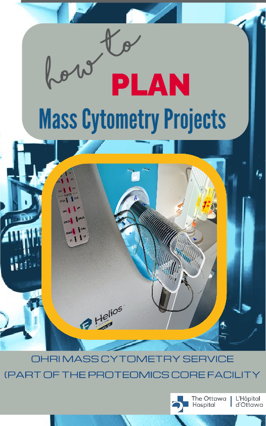
Sample Acquisition
At OHRI, we acquire samples on a 3rd-generation Helios instrument with top-of-the-line sensitivity and the highest mass range available. If you're more used to flow cytometry there are differences that you should be aware of:
- 500 events / second acquisition rate
- Samples are acquired in ddH2O
Data Analysis
For mass cytometry we rarely use manual gating techniques. Most often we use high parameter workflows consisting of:
- Dimensionality Reduction
- e.g. tSNE, hSNE, UMAP
- Clustering
- e.g. Phenograph, FlowSOM, SPADE
- Trajectory Analysis
- e.g. Pseudotime
We're here to help
The information provided here is a very brief introduction to many of the things you should be thinking about when planning a mass cytometry project. We appreciate that these are complex topics. If you have any questions about any aspect, let us know. We're here to help! Email us at dcarragher@ohri.ca with any queries